The Joint Between the L2 and L3 Vertebrae Is a
For example a L2-L5 anterior fusion requires the assignment of only one fusion code with the body part being 1. The L3 vertebra is in the middle of the five 5 lumbar vertebrae in the lower back portion of the spinal column.

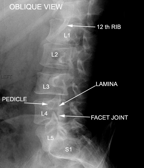
Pin On Health
Part of the L4 root joins with L5 to form the lumbosacral trunk which then joins the sacral plexus.

. TheFacet L2-L3 disc is about 2 inches above the waist. However a L2-S1 anterior fusion requires two fusion codes with one code being. The C6 vertebra is located in the inferior end of the neck just above the thorax.
Problems at the L2. The general characteristics of the third through sixth cervical vertebrae are described here. The L2-L3 spinal segment requires a specialized set of skills and methods for complete recovery with lasting effects.
The abdominal aorta is a continuation of the thoracic aorta beginning at the level of the T12 vertebrae. It is found in the base of the neck between the C5 and the last cervical vertebra C7. Anterior rami of spinal nerves L1-L3 and femoral nerve L2-L4 iliacus only Gluteal muscles superficial Muscles.
Key facts about hip muscles. By convention the cervical vertebrae are numbered with the first one C1 closest to the skull and higher numbered vertebrae C2C7 proceeding away from the skull and down the spine. Because of the substantial weight-bearing role of the L3 a role it shares with all the lumbar vertebrae this bones vertebral arch and cylindrical centrum vertebral body are massively built-among the largest of the moveable vertebrae.
Have You Been Diagnosed with Slip-Disc At L2-L3. At this level the aorta terminates by bifurcating into the right and left common iliac arteries that supply the lower body. The main nerves formed by the plexus are the femoral nerve the obturator nerve and the lateral femoral cutaneous nerve.
The anatomy of the lumbar spine is quite complex. Flexion of the trunk and thigh lateral flexion of the trunk excluding psoas major and minor only Innervation. The lumbar spine makes up the the lower end of the spinal column.
The lumbar plexus in the human arises from T12 L1 L2 L3 and L4 spinal nerves. Iliacus psoas major and psoas minor Main function. Here we will attempt to provide a brief overview of lumbar spinal anatomy.
The first second and seventh vertebrae are extraordinary and are detailed later. A disc made of a soft gelatinous core nucleus pulposus surrounded by tough layers of fibrous tissue annulus fibrosus is situated between the. The L3 and L4 vertebrae are connected at the back by a pair of facet joints zygapophyseal joints which are covered by articulating cartilage to provide smooth movements between the joint surfaces.
Gluteus maximus gluteus medius. Slip-disc at L2-L3 is descriptive of a spinal disc disorder of the upper lumbar spine. Paired parietal arteries arising.
It consists of 5 lumbar vertebra that are numbered 1 through 5 from top to bottom ie. It is approximately 13cm long and ends at the level of the L4 vertebra. L1 L2 L3 L4 and L5.
Contact one of our centers near you today. It is the second most. The intervertebral joint is the space that is located between any two adjacent vertebrae.
The C6 vertebra plays an important role in supporting and protecting the structures of the head and neck as well as anchoring the muscles that move and support the neck. One factor in determining the number of fusion codes to assign is how many levels were fused.

Anatomy Bones Medical Anatomy Human Skeleton Anatomy

Pediagenosis Spinal Nerve Lumbar Vertebrae

Pin On Sciatica

Pin On Skelet

Mri Spine Anatomy Free Mri Lumbar Spine Sagittal Cross Sectional Anatomy Anatomy Images Mri Spines

Nerves Of The Lumbar Spine Medical Anatomy Human Anatomy And Physiology Plexus Products

Pin On Fascia First Chiropractic

Pin On Fit

Pin On Medial Branch Block

Pin On Back Injuries Spine Disorders What S Causing My Back Pain Lower Middle Upper Back Problems

Pin On Disorders

Pin On Projects To Try

The Vertebral Column In Lateral View Showing The Curvatures Medical Anatomy Human Bones Anatomy Human Anatomy And Physiology
Large Color Medical Illustration Shows The Diaphragm And Phrenic Nerves With Nearby Anatomical Structures Clear Human Anatomy And Physiology Nerve Respiratory
Radiographic Anatomy Of The Skeleton Cervical Spine Anteroposterior Ap View Labelled Radiology Imaging Radiology Medical Anatomy

Pin On Anatomy And Physical Therapy

Pin On Lumbar Spinal Stenosis

Degeneration Of L4 L5 And L5 S1 Intervertebral Discs Intervertebral Disc Degenerative Disc Degenerative Disc Disease

Shsmnyfvhutzxm


Comments
Post a Comment