How Does Bright Field Microscopy Allow Images to Be Visualized
The name brightfield is derived from the fact that the specimen is dark and contrasted by the surrounding bright viewing field. The bright-field microscopy produces.

Low Cost Fluorescence And Brightfield Microscopes Microscopic Fluorescence Microscopy Microscopy
The background will appear more radiant in bright field microscopy.

. The bright-field microscope is your standard microscope that you could purchase for your niece or nephew at any toy store. What type of microscope would you use to view a whole earthworm. Phase contrast microscopy allows scientists to visualize specific organelles directly under a microscope without staining or otherwise contaminating the specimen.
Recently while recording an image in confocal microscopy i found that if Z sectioning of a 10 micrometer fluorescence bead is done it looks like 15 micrometer in the Z. Therefore the tested organism that has absorbed the part of the light will appear darker and the remaining ie. Specimens are viewed under phased light to improve magnification.
Who are the experts. A light field microscope is created by inserting a microlens array D of focal length at the intermediate image plane of an optical microscope consisting of an objective B with focal length and tube lens T with focal length The sensor E is now placed at the back focal plane of the microlens array. A microscope is primarily used to enlarge or magnify the object that is being viewed which can not otherwise be seen by the naked eye.
--Specimens are fixed and have bright fluorescent molecules attached to them. Concentration of light rays coming from the object 2. The bright-field microscope magnifies or enlarges an object so that it is visible to the observer.
Bright-field microscopy uses light from the lamp source to illuminate the specimen. The degree of fineness with which an image can be recorded or produced often expressed as the number of pixels per unit of length typically an inch. Electron microscopes allow for higher magnification in comparison to a light microscope thus allowing for visualization of cell internal structures.
Brightfield microscopy is the most elementary form of microscope illumination techniques and is generally used with compound microscopes. Bright-field microscopy is a simple method to perform. The different ray bundle diagrams correspond to paraxial rays from points.
Detection of different organisms present in the same specimen. A brightfield microscope is basically any type of light microscope that makes use of an illumination technique called bright field microscopy. Bright field microscopy is the conventional technique.
Can virus be seen with a light microscope. Magnification is achieved using a two-lens system composed of the ocular lens and the objective lens. How does brightfield microscopy allow images to be visualized.
There are several different techniques for transmitted light that are used for sample analysis. The main functions of the objective lenses are -1. Forming the image of the specimen and 3 Magnifying the image.
Brightfield Microscopy Uses. Specimens are illuminated with white light. The name brightfield is derived from the fact that the specimen is dark and contrasted by the surrounding bright viewing field.
Produce a dark image against a brighter background. Most bright-field microscopes are equipped with three. Because of the hands off approach that phase contrast allows scientists have been able to observe cellular functions and processes that many phase object microorganisms undergo that were previously very difficult.
The limitations of bright-field microscopy include low contrast for weakly absorbing samples and low resolution due to the blurry. The lens at the top that you look through. Images produced with brightfield illumination appear dark andor highly colored against a bright often light gray or white background.
This digital image gallery explores a variety of stained specimens captured with an Olympus BX51 microscope coupled to a 12-bit QImaging Retiga camera system and a three-color liquid crystal tunable filter. --Specimens are illuminated with blue light to visualize internal features of cells. Bright-field microscopy is one of the simplest optical microscopy.
Electrons strike the specimen being examined. Rapid preliminary organism identification. An earthworms internal organs can be viewed through a dissecting microscope.
This light is gathered in the condenser then shaped into a cone where the apex is focused on the plane of the specimen. This kind of microscope has its own light source that provides illumination to the specimen as well as at least one lens to magnify the image of that specimen. Among them electron microscopy EM has played a major role due to the small size of virus particles that with very few exceptions cannot be visualized by conventional light microscopy 1234.
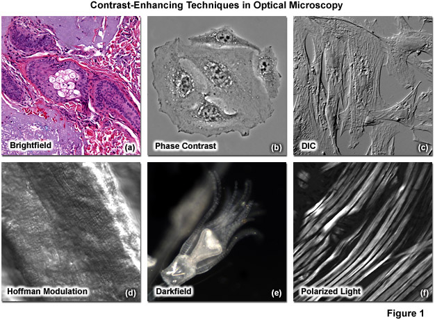
Experts are tested by Chegg as specialists in their subject area. Polarized light microscopy is a contrast-enhancing technique that dramatically improves the quality of an image acquired with birefringent materials when compared to other techniques such as brightfield and darkfield illumination phase contrast differential interference contrast fluorescence and Hoffman modulation contrast. How does brightfield microscopy allow images to be visualized.
Use of a microscope to magnify objects too small to be visualized with the naked eye so that their characteristics are readily observable. It can quickly produce a magnified image of the fixed specimens and live cells. Specimens are fixed and have bright fluorescent molecules attached to them.
This was achieved through so-called computer-enhanced bright-field imaging which involves averaging over multiple frames background subtraction spatial. They are usually 10X or 15X power. Key Terms resolution.
Rapid final identification of certain organisms. Pros and Cons Brightfield microscopy is the most elementary form of microscope illumination techniques and is generally used with compound microscopes. --Specimens are viewed under.
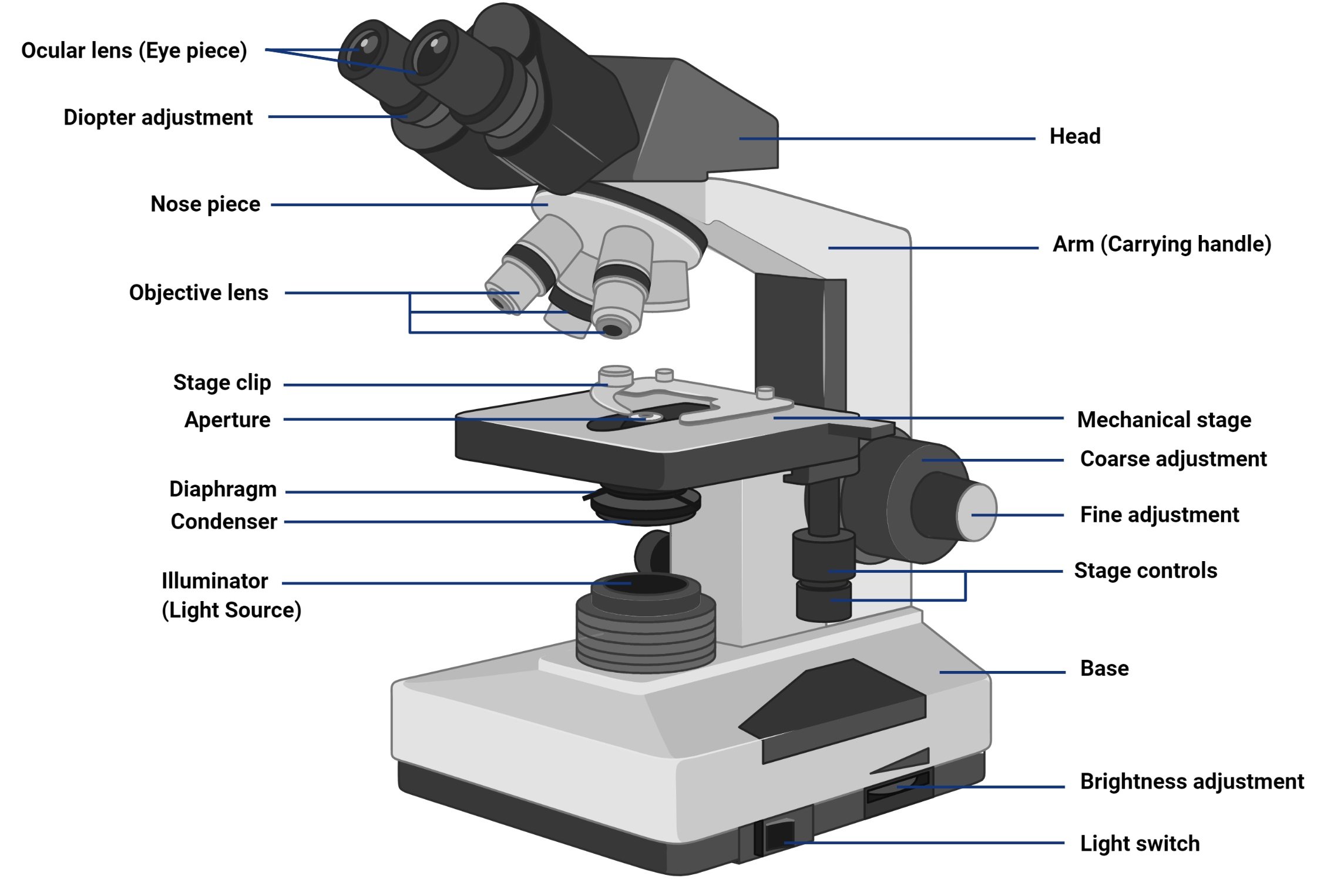
Here is a website on the basic parts of a bright-field compound microscope in case you are not enrolled in the general microbiology lab. What lens do you look through first on a microscope. The specimen is illuminated by a light source at the base of the microscope and then initially.
How does brightfield microscopy allow images to be visualized. In bright-field microscopy illumination light is transmitted through the sample and the contrast is generated by the absorption of light in dense areas of the specimen. What microscope uses bright field microscopy.
What Is Bright Field Microscopy Quora
What Is A Microscopic Technique Which Allows For The Visualization Of Live Unstained Cells Quora
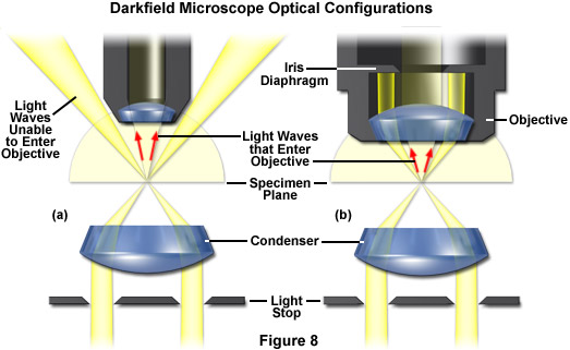
Zeiss Microscopy Online Campus Microscopy Basics Enhancing Contrast In Transmitted Light

Bright Field Microscopy Conduct Science

Brightfield An Overview Sciencedirect Topics
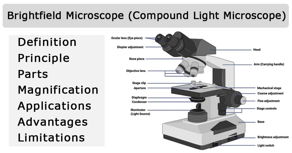
Brightfield Microscope Compound Light Microscope Definition Principle Parts
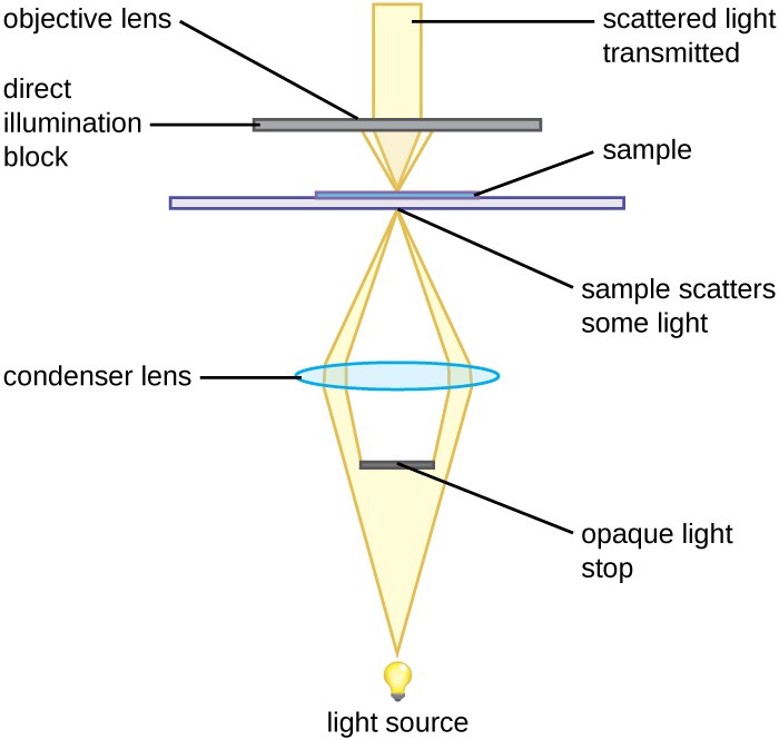
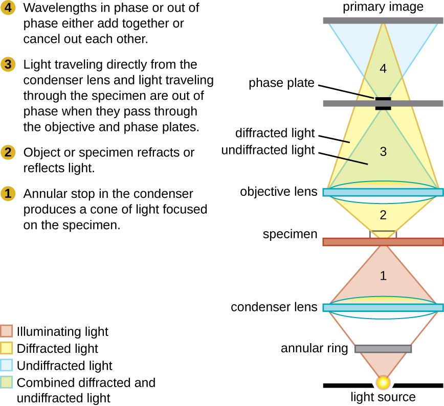
Instruments Of Microscopy Microbiology
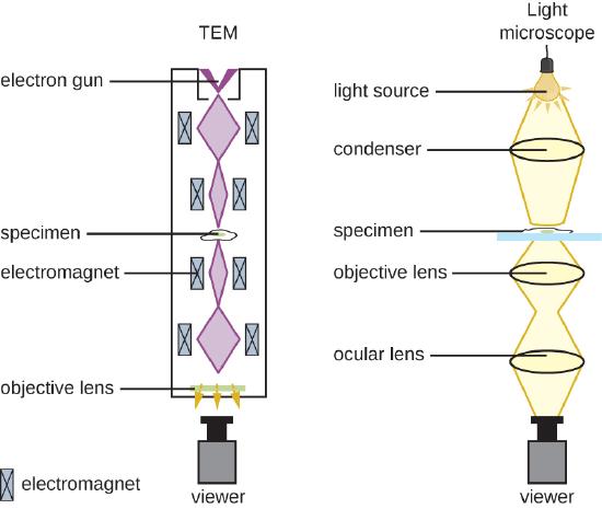
2 3 Instruments Of Microscopy Biology Libretexts
Brightfield Microscope Compound Light Microscope Definition Principle Parts

A I Bright Field Microscopy Image Of A Monolayer Of Metal Coated Download Scientific Diagram

Scanning Electrochemical Cell Microscopy In A Glovebox Structure Activity Correlations In The Early Stages Of Solid Electrolyte Interphase Formation On Graphite Martin Yerga 2021 Chemelectrochem Wiley Online Library
What Is Bright Field Microscopy Quora

Cygel By Biostatus Limited Microscopy Gaming Logos Reading
What Is Bright Field Microscopy Quora
What Is Bright Field Microscopy Quora

Brightfield An Overview Sciencedirect Topics
Instruments Of Microscopy Microbiology
Zeiss Microscopy Online Campus Microscopy Basics Enhancing Contrast In Transmitted Light

Coherent Anti Stokes Raman Scattering Microscopy For Polymers Xu Journal Of Polymer Science Wiley Online Library
Comments
Post a Comment